Books by New York TImes Bestselling Author Robert Lufkin

LIES I TAUGHT IN MEDICAL SCHOOL
Based on Dr Lufkin’s experience as a full professor at both UCLA and USC medical schools. The book is a riveting, cautionary tale of how medicine has gotten things so wrong (and continues to) in several key areas:
-how chronic diseases are all linked by common root causes overlooked by our system.
-how financial incentives, simple human error, and other factors drive the soaring rates of chronic disease.
-how Dr. Lufkin was able to reverse these diseases in himself by changes in lifestyle that anyone can do.
The book provides detailed instructions on how to keep these errors from ruining your health.
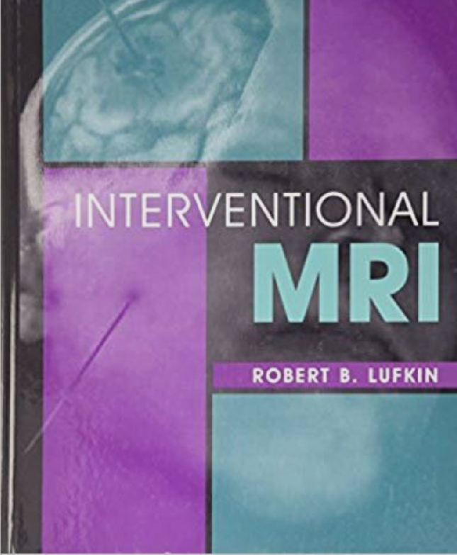
Interventional MRI
In this first-ever volume, INTERVENTIONAL MRI presents a comprehensive, up-to-date assessment of the new, rapidly growing field of MR-guided therapy. Lavishly illustrated with nearly 550 images, it provides in-depth, state-of-the-art coverage of instrumentation, techniques, and clinical applications. Parts I and II cover instrumentation and general interventional MR guidance techniques. Part III covers image-guided treatment techniques, and part IV covers clinical applications. Provides the first-ever comprehensive, up-to-date overview and state-of-the-art assessment of interventional MRI for a wealth of general information on this emerging field. Includes the most current information on instrumentation, techniques, and clinical applications, as well as economics, start-up, and database management issues. Features 548 state-of-the-art images, including 44 in color, to visually support and enhance the text. Includes contributions from an international cast of more than 90 leaders in the field for the most up-to-date and reliable information.
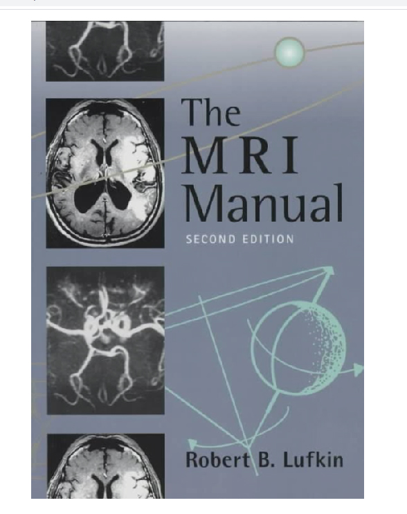
The MRI Manual
Extensively rewritten and expanded, this new second edition remains the most thorough, practical, and economical introductory guide to both the principles and the clinical applications of MRI. It offers comprehensive yet concise coverage of all aspects of MR physics and technology, plus complete discussions of clinical aspects — all with an informal and direct writing style for ease of use. A conveniently-sized paperback, it serves triple duty as an introductory text, a day-to-day manual, and a handy reference for those new to the field of MR, as well as for those who need an update on recent developments in technology and practice.
Includes more than 600 high-quality illustrations and MR scans to enhance descriptions of clinical aspects of MRI. Highlights the effects of MR physics and technology on imaging throughout — a topic often left uncovered in most introductory MR texts. Describes normal MR appearances of organs at different pulse sequences for ease of comparison. Presents imaging strategies and protocols for each disease process, including recommended pulse sequences, use of surface coils, and MR contrast agents. 27077 Provides a comparative look at the diagnostic advantages and disadvantages of MR versus other modalities in similar situations. Offers complete coverage of MR technique and preparation, with special emphasis on the pediatric patient Discusses the role of MRI in staging tumors, assisting the reader with this often difficult process.
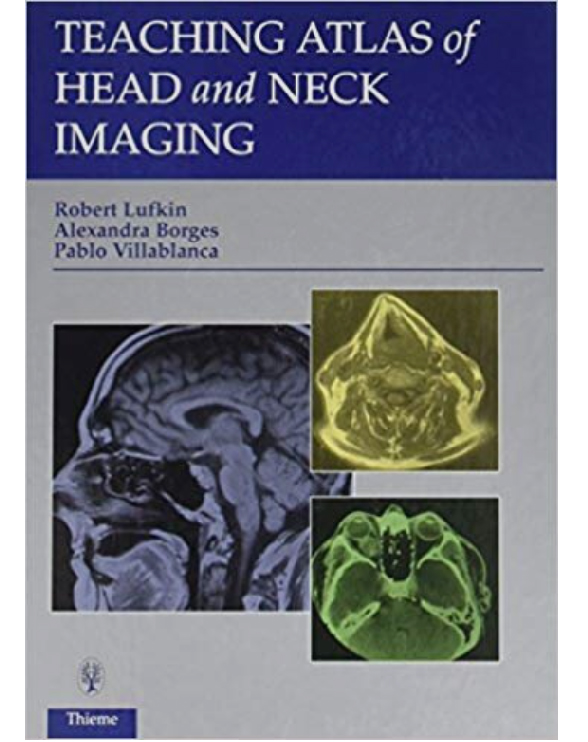
Teaching Atlas of Head and Neck
Devoted specifically to the complex region of the head and neck, this clinically oriented book brings you up-to-date on new imaging protocols and patient strategies. You’ll find complete coverage of all imaging modalities, including their advantages and disadvantages in obtaining a complete work-up of the patient. More than 100 cases stress “real-life” clinical problems, supported by 700 high-quality radiographs and illustrations.
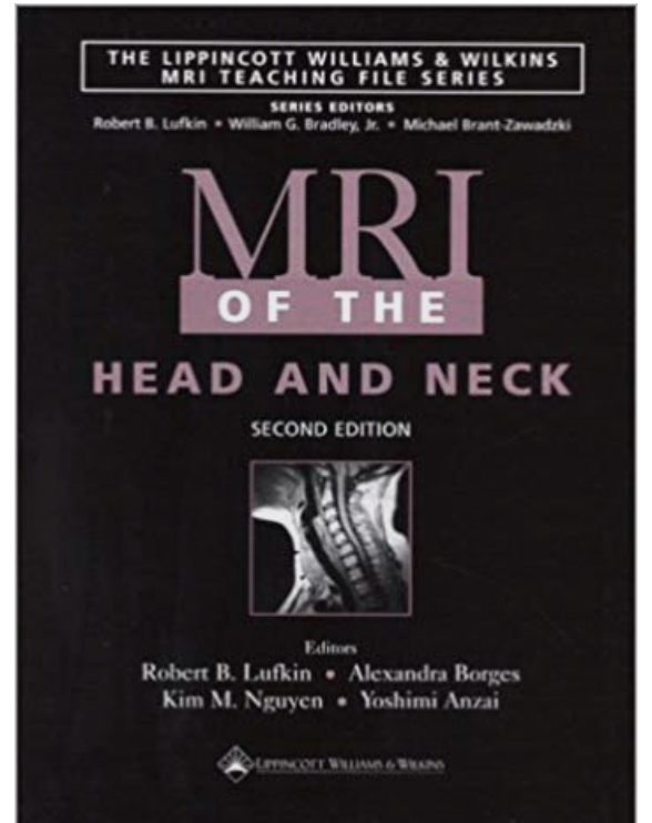
MRI of the Head and Neck
The thoroughly revised and updated Second Edition of this text is part of the popular Lippincott Williams & Wilkins MRI Teaching File Series. The book presents 100 actual case studies that cover a wide range of head and neck disorders and demonstrate the use of current MRI techniques and contrast enhancement agents to aid in diagnosis. Each case study is illustrated with high-resolution MR images and presented in an easy-to-follow format on a two-page spread. On the left-hand page are the images and the clinical history. On the right-hand page are concise descriptions of the radiographic findings, the diagnosis, and the pathology. This format is ideal for teaching readers how to interpret MR images or for everyday reference at the view box. Praise for the previous edition: “The book is packed with pearls…It should be useful to residents and generalists in radiology and to those in other disciplines in which MR imaging is used…This book is well worth the investment.”–American Journal of Roentgenology
Imaging of the Larynx
Not withstanding the important role of direct clinical and endoscopic examination in modern management of pathological conditions of the larynx, radiological study the and, more specifically, cross-sectional imaging by CT and MRI make definite diagnostic contributions by virtue of their potential to display superbly the deeper extent of laryngeal lesions. Indeed, remarkable progress has been achieved during recent years in CT and MRI techniques as applied to the neck region. This book sets out to provide a sorely needed update of our knowledge of the diagnostic potential of these cross-sectional methods and constitutes a very welcome addition to our series “Medical Radiology”, which aims to cover all important clinical imaging fields of modern diagnostic radiology. It will be of great interest to general and head and neck radiologists as well as to ENT surgeons and radiotherapists. Professor R. Hermans and the other distinguished contributors to this work are internationally renowned experts in the field and they have accumulated vast experience and a wealth of radio-pathological knowledge of the larynx over the years. I would like to congratulate them most sincerely for this outstanding volume, its comprehensive contents and its superb illustrations. I hope that this book will meet with the same great success as previously published volumes in the series. I would appreciate any constructive criticism that might be offered.
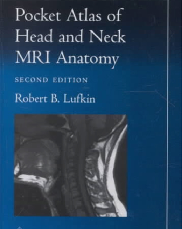
Pocket Atlas of Head and Neck MRI Anatomy 2nd Edition
The thoroughly revised Second Edition of this popular and widely used pocket atlas is a quick, handy guide to head and neck anatomy as seen in state-of-the-art magnetic resonance images. This edition presents 158 new high-resolution images of all major areas–the neck, larynx, oropharynx, tongue, nasopharynx, skull base, sinuses, temporal bone, orbit, and temporomandibular joint–displayed in axial, sagittal, and coronal planes. Anatomic landmarks on each scan are labeled with numbers that correlate to a key at the top of the page. An illustration alongside the key indicates the plane.Praise for the previous edition:”A nice introduction for practicing radiologists who are new to MR of the no-man’s land between the skull base and thoracic inlet. Imaging of the head and neck is a growing segment of many radiology practices, and familiarity with this type of normal anatomy is necessary….This is a nice and inexpensive guide to keep at hand in film-viewing areas.”–American Journal of Roentgenology
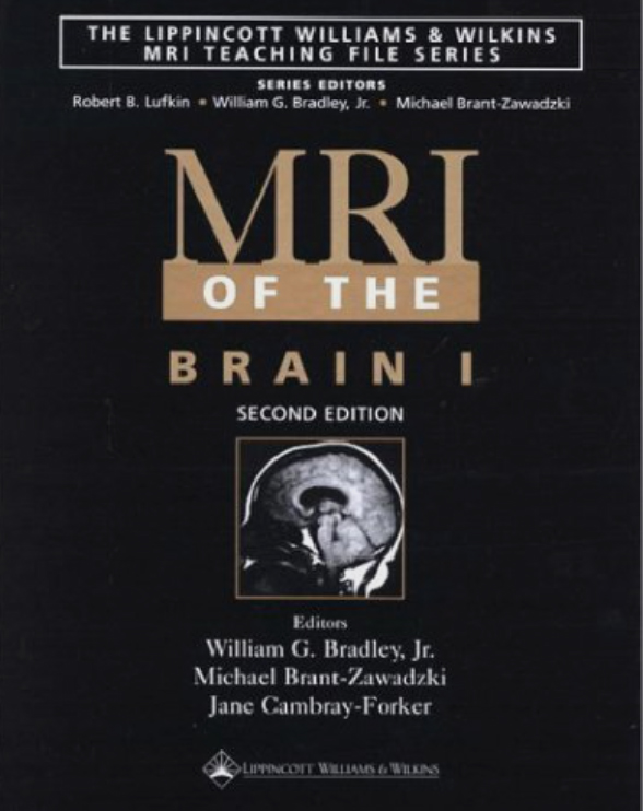
MRI of the Brain I
Extensively revised and updated for their Second Editions, MRI of the Brain 1 and MRI of the Brain 2 are part of the popular Lippincott Williams & Wilkins MRI Teaching File Series. Each volume presents 100 actual case studies illustrated with high-resolution MR images. The images were obtained with state-of-the-art scanning techniques and selected on the basis of clinical utility.Each case is presented in an easy-to-follow format on a two-page spread. On the left-hand page are the images and the clinical history. On the right-hand page are concise descriptions of the radiographic findings, the diagnosis, and the pathology. This format is ideal for teaching readers how to interpret MR images or for everyday reference.The cases in MRI of the Brain 1, Second Edition cover flow phenomena, vascular abnormalities, hemorrhage, trauma, and more. MRI of the Brain 2, Second Edition includes hydrocephalus, infarction, and demyelinating disease. Together, these two volumes cover a wide array of diseases of the brain, including both non-neoplastic diseases and neoplasms.Praise for the previous edition:”The text is presented in a way that simulates good clinical teaching at the view box….The format is clearly more effective than that of the usual atlas, and the book will be an excellent companion for the traditional neuroradiology textbooks.”–American Journal of Neuroradiology
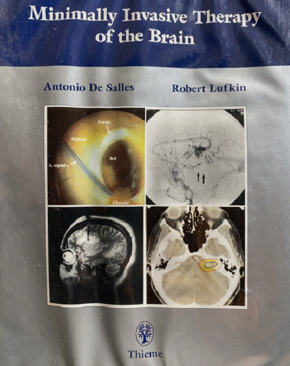
Minimally Invasive Therapy of the Brain
The latest techniques on stereotaxis & brain mapping, image-guided interventional radiology, & neuroendoscopy are discussed by an international group of neurosurgeons, radiologists, & radiation oncologists.
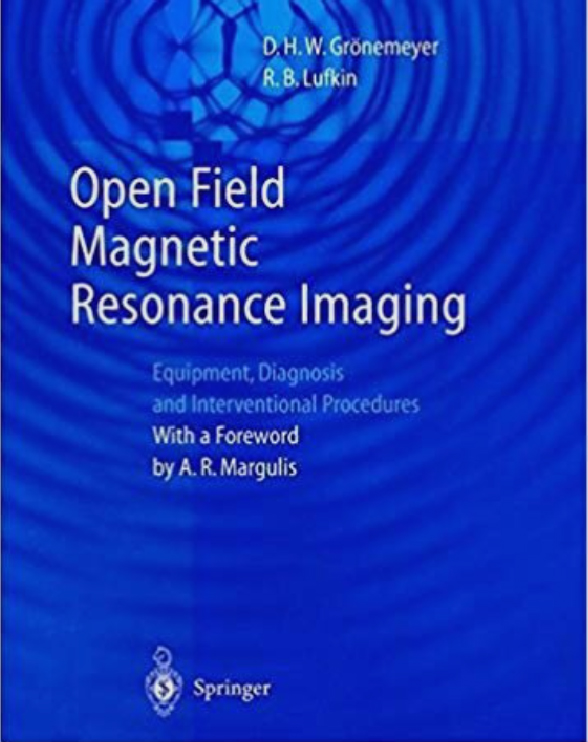
Open Field Magnetic Resonance Imaging
Covers issues related to the practice of both diagnostic and interventional MRI on this new type of open design MR scanner. The unique configuration and often mid-field strengths of these machines necessitates new strategies for both diagnostic and interventional procedures compared to that ofthe standard 1.5 tesla diagnostic-only MR scanners, to which the majority of the radiologic literature is addressed today. This broad, multi-authored work will appeal to radiologists, medical physicists, other physicians, and health care personnel
MR Imaging and Spectroscopy of Central Nervous System Infection
Central nervous system (CNS) infections continue to pose a serious problem in health care even with improved knowledge and treatment. Despite the introduction of newer antimicrobial agents and diagnostic techniques, the morbidity and mortality associated with CNS infections remain high. The morbidity associated with CNS infections may be even more important than the death rate especially in developing countries as neurological sequelae may deprive the survivors of intellect and physical ability, demeaning the quality of life and burdening health resources and social services. MR imaging is an important integral part of the protocol for the management of CNS infections and MR spectroscopy is increasingly being utilized in its management. This work is an attempt to provide a comprehensive review of imaging and spectroscopy of the commonly encountered CNS infections in the clinical practice in developing and developed countries. The first chapter deals with basic physical principles of MR imaging and spectroscopy that will help beginners to understand the technical terms used in subsequent chapters. The remaining 10 chapters deal with clinical, pathological, MR imaging and spectroscopy features and their applications in CNS infections. This will help in giving a comprehensive understanding to readers with a background in clinical, radiological, basic MRI, and neurological sciences. The T2 hypointense lesions are a real diagnostic dilemma especially in developing countries for which an algorithm has been suggested in the concluding chapter.
MRI of the Head and Neck (MRI TEACHING FILE)
This book presents individually bound guides for each section of the body, the 1,000 concise and clearly illustrated case files cover neoplastic, non-neoplastic, degenerative, inflammatory, congenital, and acquired disease of the brain, head and neck, spine, musculoskeletal system, cardiovascular system, chest, abdomen, and male and female pelvis. It focuses on specific body regions; one is devoted to pediatric MRI; and one reviews the principles of MRI and identifies frequently encountered artifacts. It contains 100 completed case studies, with high-resolution MR images.
MRI: Principles and Artifacts
MRI: Principles and Artifacts is the perfect resource for experienced MR users looking to build on their understanding of the techniques, technology, and capabilities of magnetic resonance imaging. Using the uniquely effective Raven MRI Teaching File format–100 case studies illustrated with MR images–the book demonstrates the practical imaging options in MR and addresses the decision-making problems arising in the day-to-day use of MR. The case studies show how to put basic MR principles to work, how to choose among different imaging options, and how to determine and correct the causes of artifacts. Forty case studies show the MR user what imaging options are available and how imaging choices affect image quality. Many of these case studies deal with newer techniques such as fast spin echo, gradient echo, magnetization transfer, diffusion, STIR, and FLAIR imaging. Sixty case studies illustrate and explain common artifacts and imaging problems in MRI and magnetic resonance angiography.
Pediatric MRI
This volume of the Raven MRI Teaching File set is a uniquely practical guide to interpreting magnetic resonance images of disorders seen in neonates, infants, and children. It contains 100 case studies illustrated with high-resolution MR images. The concise text that accompanies each case study and set of images provides all essential information on clinical history, radiographic findings, and diagnosis and explains how the diagnosis was reached.
MRI of the Spine
This volume of the Raven MRI Teaching File set is a uniquely practical guide to interpreting magnetic resonance images of spinal disorders. It contains 100 case studies illustrated with high-resolution MR images. The concise text that accompanies each case study and set of images provides all essential information on clinical history, radiographic findings, and diagnosis and explains how the diagnosis was reached.
MRI of the Brain Volume 2
The thoroughly revised and updated Second Edition of this acclaimed text presents 100 actual case studies that show readers how to interpret MR images of brain disorders. Each case study is illustrated with high-resolution MR images and presented in an easy-to-follow format on a two-page spread. On the left-hand page are the images and the clinical history. On the right-hand page are concise descriptions of the radiographic findings, the diagnosis, and the pathology. This format–ideal for teaching or for everyday reference at the view box–is the most popular feature of The Lippincott Williams & Wilkins MRI Teaching File Series.The cases in MRI of the Brain 2, Second Edition include hydrocephalus, infarction, and demyelinating disease. MRI of the Brain 1, Second Edition covers flow phenomena, vascular abnormalities, hemorrhage, trauma, and more. Together, these two volumes provide excellent coverage of most brain diseases encountered in clinical practice, including both non-neoplastic diseases and neoplasms.Praise for the previous edition:”The book is valuable to a wide audience….This is an excellent teaching device and is worth the purchase price.”–American Journal of Roentgenology”This is a wonderful teaching file for MRI of the brain.”–Magnetic Resonance Imaging
MRI of the Cardiovascular System
This volume of the Raven MRI Teaching File set is a uniquely practical guide to interpreting magnetic resonance images of cardiovascular diseases. Illustrated with more than 600 high-resolution MR images, the book contains 100 case studies made at large referral university hospitals, covering a wide variety of diseases. The concise text that accompanies each case study provides all essential information on clinical history, radiographic findings, and diagnosis and explains how the diagnosis was reached. Coverage includes both common and unusual cases.
International Editions
RM Intervencionista (Spanish edition)
Obra de gran actualidad y vigencia al tratar un tema de importancia creciente en la sanidad actual como es la RM intervencionista, que ha pasado de ser una “gran idea o “un concepto con mucho futuro a una realidad aplicable y recomendable (no invasiva, más económica…) a una gran variedad de casos. La increíble capacidad de imagen de partes blandas de la RM hace que se le hayan buscado a esta tecnología aplicaciones terapéuticas y no sólo diagnósticas. Hoy en día encontramos aplicaciones guiadas por RM en el ámbito de la mama, cerebro, mediastino, abdomen, sistema musculoesquelético, cabeza, cuello, etc.Con una estructura en cuatro partes, en las que se revisa desde los aspectos técnicos de la RM a sus posibles aplicaciones clínicas. (Spanish edition)
Cep Atlası: Baş Boyun MR Görüntüleme Anatomisi (Turkish edition)
The thoroughly revised Second Edition of this popular and widely used pocket atlas is a quick, handy guide to head and neck anatomy as seen in state-of-the-art magnetic resonance images. This edition presents 158 new high-resolution images of all major areas–the neck, larynx, oropharynx, tongue, nasopharynx, skull base, sinuses, temporal bone, orbit, and temporomandibular joint–displayed in axial, sagittal, and coronal planes. Anatomic landmarks on each scan are labeled with numbers that correlate to a key at the top of the page. An illustration alongside the key indicates the plane.Praise for the previous edition:”A nice introduction for practicing radiologists who are new to MR of the no-man’s land between the skull base and thoracic inlet. Imaging of the head and neck is a growing segment of many radiology practices, and familiarity with this type of normal anatomy is necessary….This is a nice and inexpensive guide to keep at hand in film-viewing areas.”–American Journal of Roentgenology
Atlas of MR Anatomy of the Head and Neck [Japanese edition]
Devoted specifically to the complex region of the head and neck, this clinically oriented book brings you up-to-date on new imaging protocols and patient strategies. You’ll find complete coverage of all imaging modalities, including their advantages and disadvantages in obtaining a complete work-up of the patient. More than 100 cases stress “real-life” clinical problems, supported by 700 high-quality radiographs and illustrations.
Pocket Atlas of Head and Neck MRI Anatomy
[Mandarin edition]
The thoroughly revised Second Edition of this popular and widely used pocket atlas is a quick, handy guide to head and neck anatomy as seen in state-of-the-art magnetic resonance images. This edition presents 158 new high-resolution images of all major areas–the neck, larynx, oropharynx, tongue, nasopharynx, skull base, sinuses, temporal bone, orbit, and temporomandibular joint–displayed in axial, sagittal, and coronal planes. Anatomic landmarks on each scan are labeled with numbers that correlate to a key at the top of the page. An illustration alongside the key indicates the plane.Praise for the previous edition:”A nice introduction for practicing radiologists who are new to MR of the no-man’s land between the skull base and thoracic inlet. Imaging of the head and neck is a growing segment of many radiology practices, and familiarity with this type of normal anatomy is necessary….This is a nice and inexpensive guide to keep at hand in film-viewing areas.”–American Journal of Roentgenology
La Tomografia Computerizzata e la Risonanza Magnetica nelle Neoplasie Maligne della Laringe (Italian edition)
This Italian language mono-graph provides a useful, well-organized review of computed tomography (CT) and magnetic resonance (MR) imaging Of the normal and tumorous larynx. Normal and abnormal CT anatomy is demonstrated and discussed by means of nicely chosen, well-reproduced, and effectively annotated axial sections that are organized by the individual regions and structures. There is appropriate emphasis on imaging of potential path- ways of metastases, limits of symmetry and asymmetry, and functional imaging. La Tomografia Computerizzafa e la Risonanza Magnetica nelle Neoplasie Maligne della Laringe is a valuable book for radiologists as well as oncologists and ear, nose, and throat physicians who are learning cross-sectional imaging Of the larynx.
